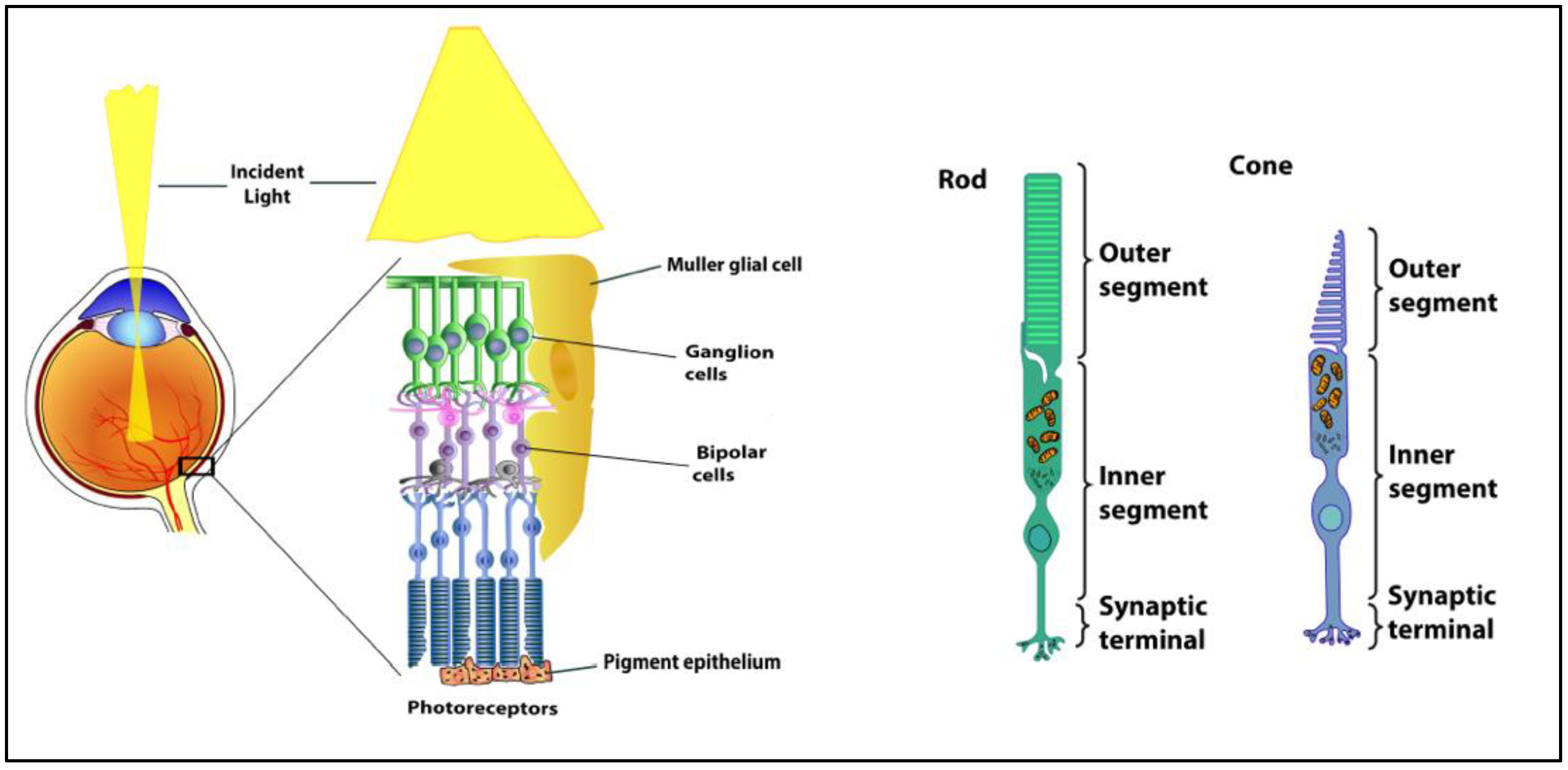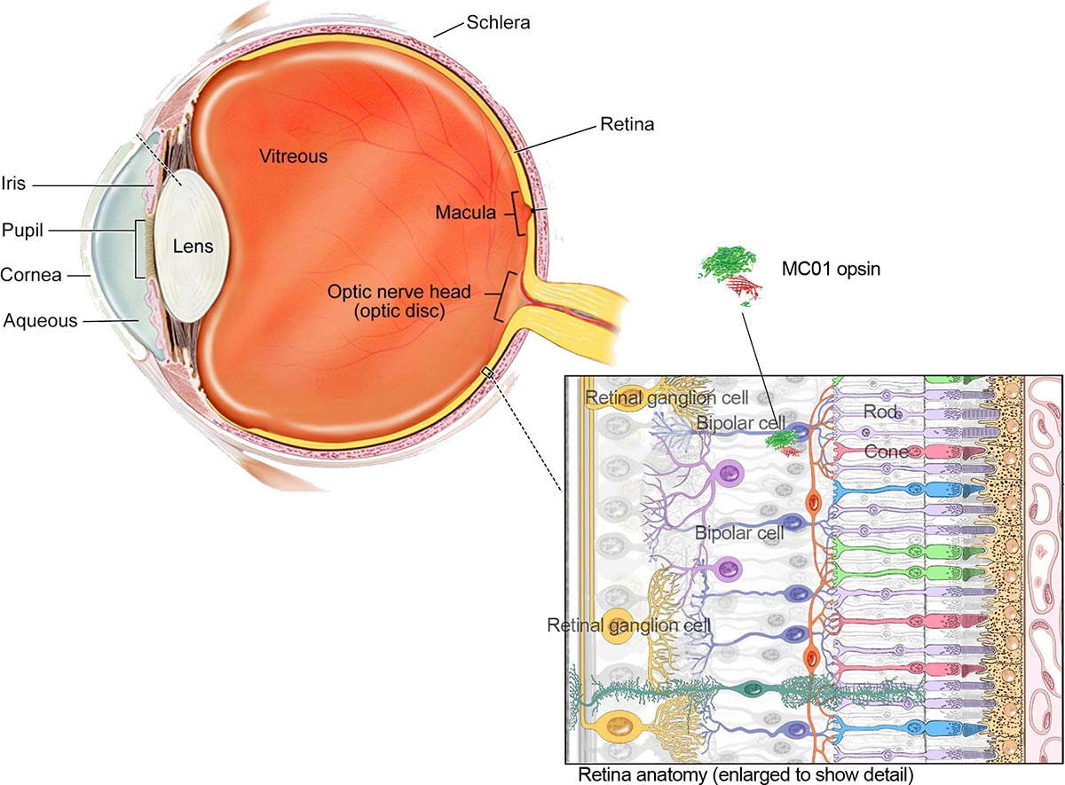

The pupil is a black dot that presents in the middle portion of the eye. The sclera delivers protection to the inner workings portion of the eye. This is a flat, white layer which is present on the outside, but the inside it is brown and comprises furrows that help the muscles of the eye to ascribe appropriately. The Sclera has generally stated the �Whites� of the Eye. The cornea also helps the eye to properly focus on light more accurately. The Cornea is basically� Eyes Outer covering.� It is generally dome-shaped layer and its main function is to protect your eye from foreign substances which may damage the Eyes inner portion.Ĭornea comprised of several layers which create a tough layer which provides additional support and protection. The eye comprised of the following parts which produce clear Vision: Eye Parts Eye Anatomy and FunctionĮye Parts and Functions are�described below in the given table. It washes the surface of the eye and stops it drying out. The tear glands produce tears, and when you blink this liquid is spread over the surface of the eye.


When you look at a bright light, the Iris Muscles work to make Pupil smaller.In the center of it is a round hole, called the Pupil. Just behind the lens is a sheet of muscle called the iris. Your brain has to correct the image so that you see it properly.The image made on the retina is upside-down. These chemical changes set off nerve impulses which travel along the optic nerve to the brain.This layer is called the retina, and light makes chemical changes in the cells of the retina. Together, the cornea and the lens behind it focus light onto a layer of sensory cells at the back of the eye.The lens is elastic and its thickness is controlled by the ciliary muscles inside – the eye.Instead, the eye focuses by changing the shape of the lens. The eyeball cannot be made longer or shorter. A camera focuses by moving the lens nearer or further away from the object.At the front of the eye is a clear, round window called the cornea.Each eye is a liquid-filled ball 2.5 cm in diameter. The eye is rather like a living Camera.For additional information visit Linking to and Using Content from MedlinePlus.Diagram of Human Eye with Labelling Eye Anatomy���Ĭomplete Physiology of Eye is described below in the given paragraph: Any duplication or distribution of the information contained herein is strictly prohibited without authorization. Links to other sites are provided for information only - they do not constitute endorsements of those other sites. A licensed physician should be consulted for diagnosis and treatment of any and all medical conditions. The information provided herein should not be used during any medical emergency or for the diagnosis or treatment of any medical condition. This site complies with the HONcode standard for trustworthy health information: verify here.

Learn more about A.D.A.M.'s editorial policy editorial process and privacy policy. is among the first to achieve this important distinction for online health information and services. follows rigorous standards of quality and accountability. is accredited by URAC, for Health Content Provider (URAC's accreditation program is an independent audit to verify that A.D.A.M. A farsighted person sees faraway objects clearly, while objects that are near are blurred.Ī.D.A.M., Inc. Farsightedness is often present from birth, but children can often tolerate moderate amounts without difficulty and most outgrow the condition. It may be caused by the eyeball being too small or the focusing power being too weak. A nearsighted person sees near objects clearly, while objects in the distance are blurred.įarsightedness is the result of the visual image being focused behind the retina rather than directly on it. For this reason, nearsightedness often develops in the rapidly growing school-aged child or teenager, and progresses during the growth years, requiring frequent changes in glasses or contact lenses. It occurs when the physical length of the eye is greater than the optical length. Nearsightedness results in blurred vision when the visual image is focused in front of the retina, rather than directly on it. A person with normal vision can see objects clearly near and faraway. Normal vision occurs when light is focused directly on the retina rather than in front or behind it.


 0 kommentar(er)
